 Dentalingo
Reference
Radiolucent vs radiopaque: dark and light on a dental radiograph
On a dental radiograph, radiolucent areas look dark and radiopaque areas look light. Here is what that means, which structures show which way, and what angulation errors do to the picture.
RadiolucentLooks dark
RadiopaqueLooks light
Source: MD0512 ¶3-24.
Why some areas look light
Where two teeth overlap, the manual describes a light area forming because the X-ray beam cannot get through the two dense surfaces.
Source: MD0512 ¶4-8b.
MD0512 was written for film, so it says “on the film”.
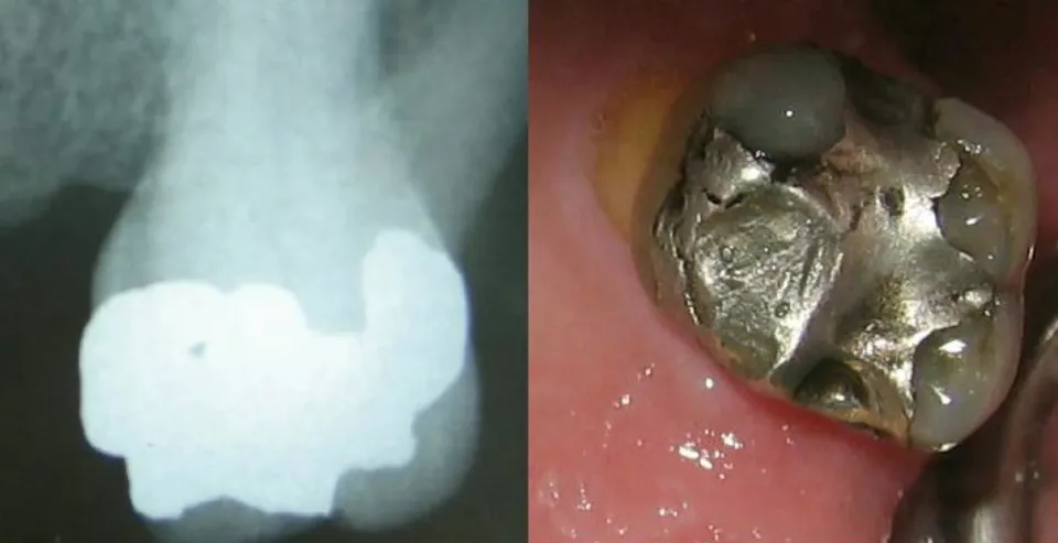

Which structures look dark, and which look light
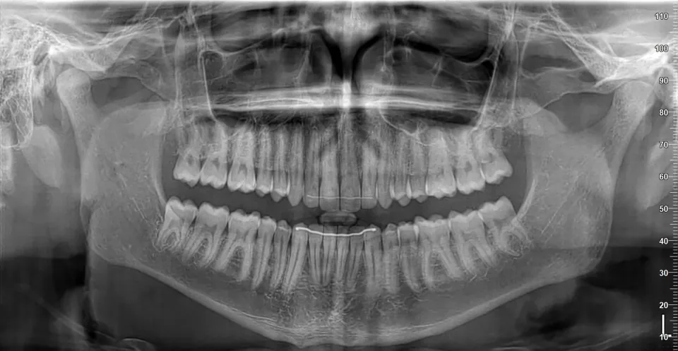

| Structure | How it looks | Source |
|---|---|---|
| Maxillary sinus | A very prominent radiolucent structure, sometimes as overlapping lobes or a single area with a radiopaque border | MD0512 ¶3-25a |
| Incisive foramen | A dark area between and above the central incisors | MD0512 ¶3-25b |
| Nasal fossae | Roughly elliptical radiolucent areas of various sizes | MD0512 ¶3-25d |
| Mandibular canal | A dark band with radiopaque borders | MD0512 ¶3-27b |
| Mental foramen | A dark area below and between the bicuspids | MD0512 ¶3-27c |
| Structure | How it looks | Source |
|---|---|---|
| Nasal septum | A radiopaque band between the nasal fossae | MD0512 ¶3-25d |
| Zygomatic arch | Commonly a well-defined radiopaque area | MD0512 ¶3-26c |
| Border of the mandible | A heavy white line | MD0512 ¶3-28a |
| External oblique ridge | A white line of variable density | MD0512 ¶3-28b |
This page lists only what its sources state. Tooth decay, the pulp, air and tooth-colored fillings are missing because none of these sources says how they look.

Angulation errors
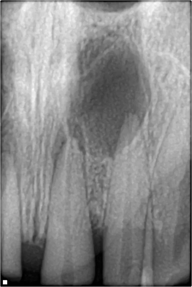
Pointing the beam at the wrong angle changes the image, in three named ways.
- Foreshortening
- The vertical angulation is larger than necessary, and the teeth look shorter than normal. (MD0512 ¶4-8a)
- Elongation
- The vertical angulation is less than necessary, and the teeth look longer than normal. (MD0512 ¶4-8a)
- Overlapping
- The horizontal angulation is incorrect, so the proximal surfaces of adjacent teeth are superimposed, leaving a light area where they overlap. (MD0512 ¶4-8b)
Practice reading radiographs
Start the free Radiation Health and Safety practice testAbout the exam
The app
Study radiography on your phone with Dentalingo
Dentalingo has 1,520 questions across the RHS®, ICE®, GC, AMP and CP exams, every answer explained and checked against a cited source, and timed mock exams with each exam’s question count and time limit.
Lessons in a path, a few minutes a day, with streaks and daily goals, spaced review of the questions you missed, and readiness by exam domain.
The first 7 questions are free. After that, full access is a Study Camp subscription; the App Store shows the price in your country. More about the app

Scan to get Dentalingo on your iPhone.
 Spaced review of what you missed
 Answers explained the moment you choose
 XP and accuracy over time
Sources
All of these are U.S. government works, in the public domain. Images are credited under each one.
- Dental Radiography (subcourse MD0512)U.S. Army Medical Department Center and School · U.S. government work, public domain