 Dentalingo
Question 1
Purpose and Technique
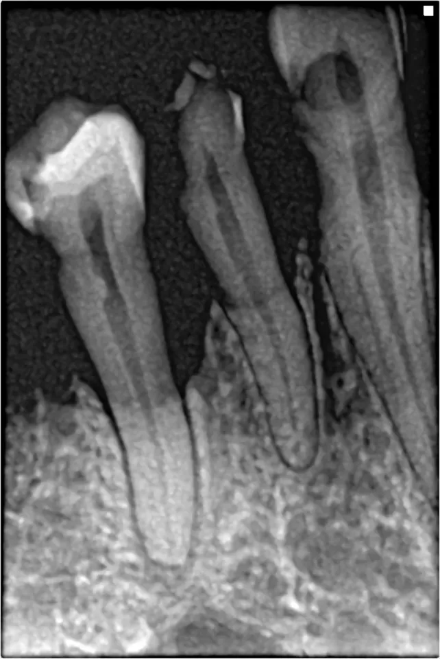
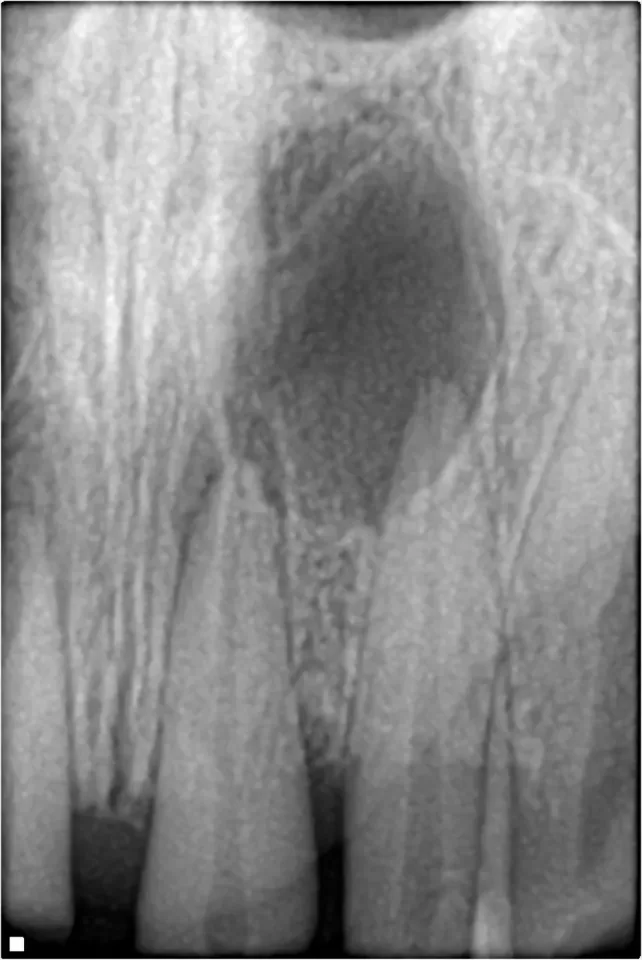
On a dental radiograph the dark and light areas are described with one pair of terms. The area visible in the bone at the root apices in this image records dark, so it is described as:
Tap an answer
Answer: B. Radiolucent, while the areas recording light on it are called radiopaque
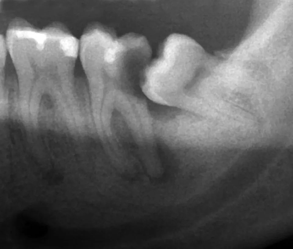
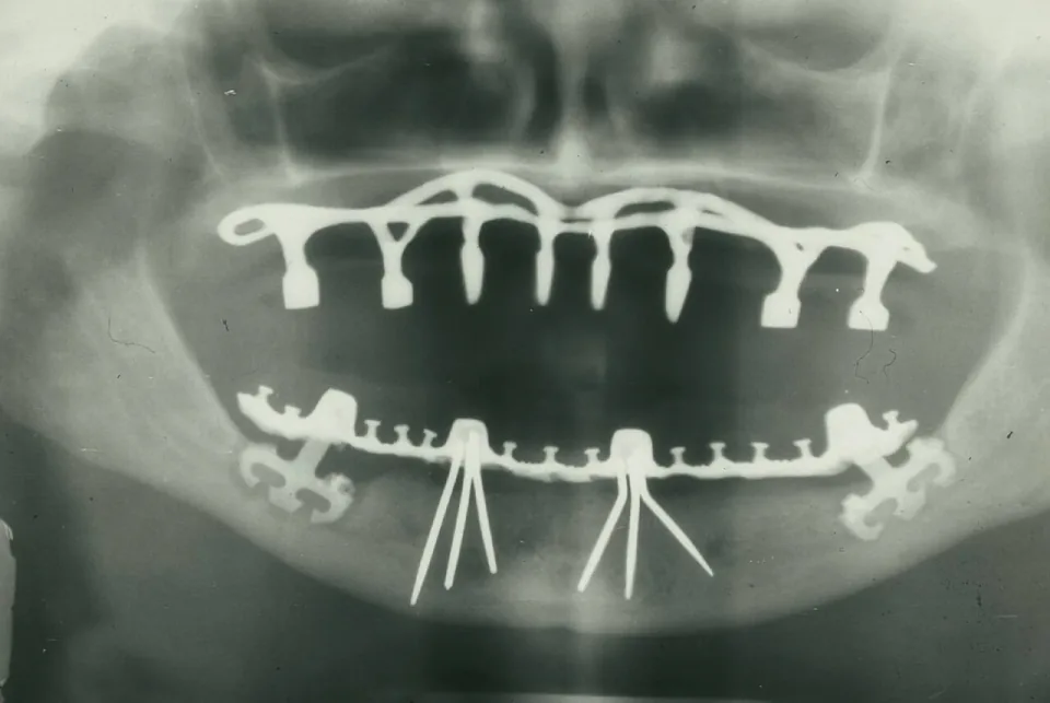
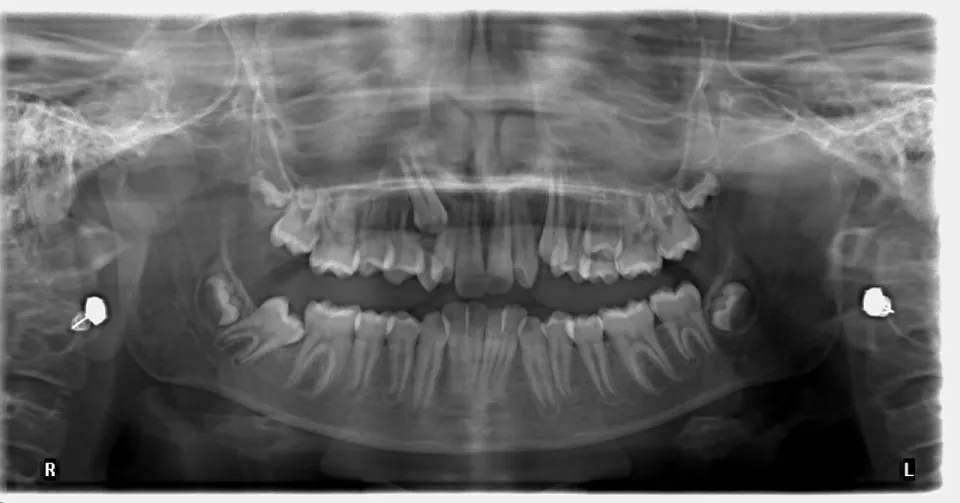
Anatomic landmarks are read by their normal appearance, and the vocabulary for that appearance is one fixed pair: the areas that record dark on a radiograph are radiolucent, and the areas that record light are radiopaque. Less dense structures such as air spaces and areas where bone is thin or absent let more of the beam reach the receptor and so record dark, while enamel, cortical bone and metal restorations absorb more of it and record light. The pair never reverses, neither term covers both appearances, and both describe the structure being imaged rather than the receptor it was recorded on.
Source: Dental Radiography (subcourse MD0512), U.S. Army Medical Department Center and School; section “3-24. GENERAL”. U.S. government work, public domain.