
Skip to content

Free practice test 2 · from 400 questions

Radiation Health and Safety practice test 2, every answer explained

Another free sample from Dentalingo’s 400 questions for the DANB® RHS® exam, none of them from practice test 1. Tap an answer to see at once whether it’s right, why, and the source it was checked against. The other 390, a timed mock exam and spaced review are in the app.

The format and domains are in the Radiation Health and Safety exam guide; how the questions are checked is on the sources page.

Question 1

Purpose and Technique

The photograph shows an intraoral x-ray tubehead with its open-ended cylinder, the position indicating device, affixed to it. What is that cylinder used for?
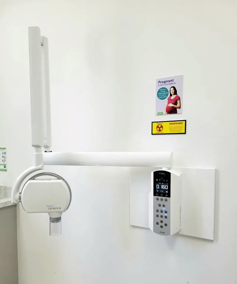
Wall-mounted intraoral x-ray unit seen across a clinic wall. A folded scissor extension arm rises up the left of the frame to a horizontal support arm, and the tubehead hangs from its far end, rotated so its branding reads upside down. A short open-ended cylinder is fixed beneath the tubehead, pointing at the floor. On the right, a separate wall-mounted control panel with a numeric display reading 0.160 and a keypad of tooth-region icons is screwed to a backing board; above it hang a 'Pregnant or think you might be' patient poster and a yellow 'Caution Radiation - Warning' placard. Nothing in the photograph is arrowed or labelled.
Image: Kiran891 · CC BY-SA 4.0 · source

Tap an answer

Answer: C. It aligns the tubehead with the patient and with the receptor that records the image

The cylinder, also called the cone or position indicating device, is affixed to the tubehead and is what aligns the tubehead with the patient and the receptor, so the beam arrives where the image is being recorded. The angulation scales that show how far the head is tilted are on the sides of the tubehead itself. Exposure settings are selected on the control panel, and holding the receptor parallel to the long axis of the tooth is the job of a receptor holder, not of the cylinder.

Source: Dental Technician, Volume 2 (NAVEDTRA 14275), U.S. Navy, 1999; section “CYLINDER”. U.S. government work, public domain.
Question 2

Purpose and Technique

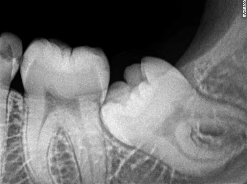
An impacted lower third molar was spotted on a panoramic image, and the dentist then requested this digital periapical of the same area. What does the intraoral periapical add beyond the panoramic view?

Digital periapical radiograph of an impacted lower third molar
Image: Nizil Shah · CC BY-SA 4.0 · source

Tap an answer

Answer: B. Sharper, higher-resolution detail of the tooth and the bone immediately around it

An intraoral receptor sits directly against the teeth, and that short object-to-receptor distance gives a periapical far finer detail than a panoramic survey, whose image is assembled through a moving slit as the tubehead travels. The added detail is what lets the dentist study the root form, the follicle and the bone immediately around an impacted molar. The panoramic had already supplied the broad view, true three-dimensional localization needs a cone-beam scan, and every extra image means an extra exposure for the patient.

Source: Dental Radiography (subcourse MD0512), U.S. Army Medical Department Center and School; section “4-40. DISADVANTAGES OF THE PANORAMIC RADIOGRAPH”. U.S. government work, public domain.
Question 3

Purpose and Technique

A removable partial denture with a metal framework was left in the patient's mouth during an intraoral exposure, so the appliance itself records its own dense outline over the region being examined. Which fault is this?

Tap an answer

Answer: A. Foreign object image

An object left in the mouth attenuates the beam and records its own outline over the teeth and bone the examination was ordered to show, and a partial denture framework is metal, so the outline it leaves is dense and unmistakable. The fault is named for its cause: a foreign object image, produced when dentures or other objects are in the patient's mouth during the exposure. That is why removable appliances come out before the receptor is placed. The other three faults come from the beam and the geometry rather than from an object in the mouth: a partial image is what is left when the tubehead is positioned so the beam misses part of the receptor, a blurred image follows movement of the patient or the tubehead while the exposure is being made, and an elongated image comes from incorrect vertical angulation.

Source: Dental Technician, Volume 2 (NAVEDTRA 14275), U.S. Navy, 1999; section “FAULTY RADIOGRAPHS”. U.S. government work, public domain.
Question 4

Purpose and Technique

A blurred image with doubled outlines of every structure most likely resulted from:

Tap an answer

Answer: D. Movement of the patient, or a drifting tubehead, while the exposure was made

Motion while the beam is on smears the projection across the receptor, so every structure records twice or as a general blur rather than with crisp margins; a patient who shifts and a tubehead that drifts produce the same picture. Prevention is a clear hold-still instruction, the shortest workable exposure time, and confirming the arm stays where it was set before pressing the switch. Approved disinfectant wipes and barriers have no effect on projection geometry, correct paralleling yields sharper images rather than doubled ones, and post-exposure sharpening alters edge contrast without duplicating anatomy.

Source: Dental Radiography (subcourse MD0512), U.S. Army Medical Department Center and School; section “3-12. BLURRED IMAGE”. U.S. government work, public domain.
Question 5

Purpose and Technique

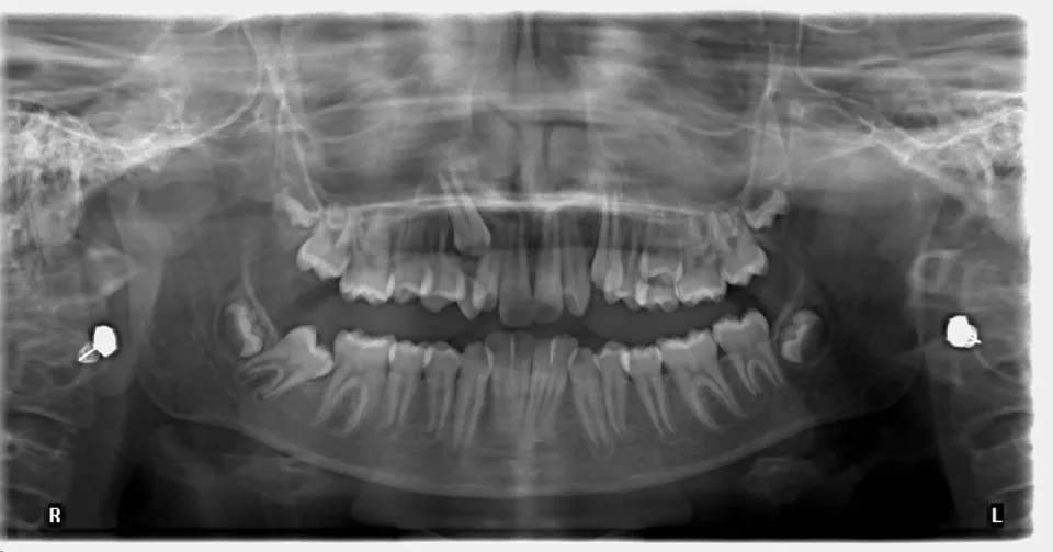
In this panoramic radiograph a heavy white line follows the very bottom outline of the lower jaw, below the roots of every lower tooth. Which landmark is that line?

Panoramic radiograph of an adult with both arches in view. On the patient's right an upper canine has not erupted: it lies in the bone with its crown pointing down and forward, overlapping the roots of the upper teeth beside it. Both lower third molars are unerupted and tilted forward. A dense bright line follows the bottom outline of the lower jaw from one side to the other. A small dense metallic object records on each side beyond the jaws at ear level, and orientation letters R and L sit in the lower corners.
Image: Challiyan · CC BY-SA 4.0 · source

Tap an answer

Answer: B. The border of the mandible, a line that has no counterpart on maxillary radiographs

The heavy white line that follows the lowest outline of the bone from one side to the other is the border of the mandible, and no similar line appears on maxillary radiographs. The other three choices are also landmarks of the lower jaw, but none of them runs along that outline. The external oblique ridge is a white line of variable density that continues forward into the molar region from the anterior border of the ramus. The mylohyoid ridge extends from close to the lower border of the symphysis upward and backward, ending beyond the third molar. The genial tubercles are round white areas with dark centers, below and between the central incisors.

Source: Dental Radiography (subcourse MD0512), U.S. Army Medical Department Center and School; section “3-28. RADIOPAQUE LANDMARKS ON MANDIBULAR RADIOGRAPHS”. U.S. government work, public domain.

Question 6

Radiation Characteristics and Protection

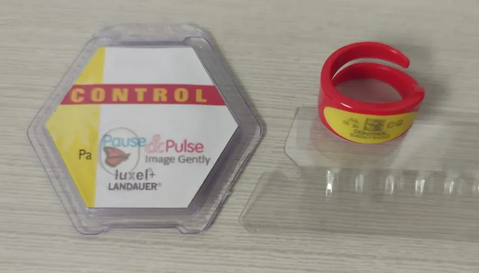
Personnel monitoring devices of this type are assigned to individual workers - the two shown are unworn CONTROL reference badges, kept away from radiation as the batch baseline. Under NRC rules, WHERE on the body such a device is worn matters because:

A flat personnel dosimetry badge in a clear plastic holder beside a red ring dosimeter, both labelled CONTROL, lying on a pale bench
Image: Reconrabbit · CC0 · source

Tap an answer

Answer: B. The dose assigned to the worker must represent the part of the body receiving the highest exposure

When external exposure is determined by measurement with a personal monitoring device, the assigned deep-dose equivalent must be for the part of the body receiving the highest exposure — so a badge worn in a low-exposure spot understates the dose of record. A dosimeter records exposure and never shields the wearer, which is the most common misconception about these devices. Ring dosimeters exist for the same reason at the extremities, where the hands may be the most exposed region.

Source: 10 CFR 20.1201, Occupational dose limits for adults, U.S. Nuclear Regulatory Commission (CFR 2024, govinfo); section “Occupational dose limits for adults”. U.S. government work, public domain.

Question 7

Radiation Characteristics and Protection

A dental assistant who exposes radiographs is pregnant. What does FDA's operator guidance direct?

Tap an answer

Answer: A. She should use a personal dosimeter regardless of anticipated exposure

FDA directs that pregnant dental personnel operating x-ray equipment should use personal dosimeters regardless of anticipated exposure levels - the general 1 mSv threshold that applies to other workers does not apply to them.

Source: The Selection of Patients for Dental Radiographic Examinations, U.S. Food and Drug Administration; section “OPERATOR PROTECTION”. U.S. government work, public domain.

Question 8

Radiation Characteristics and Protection

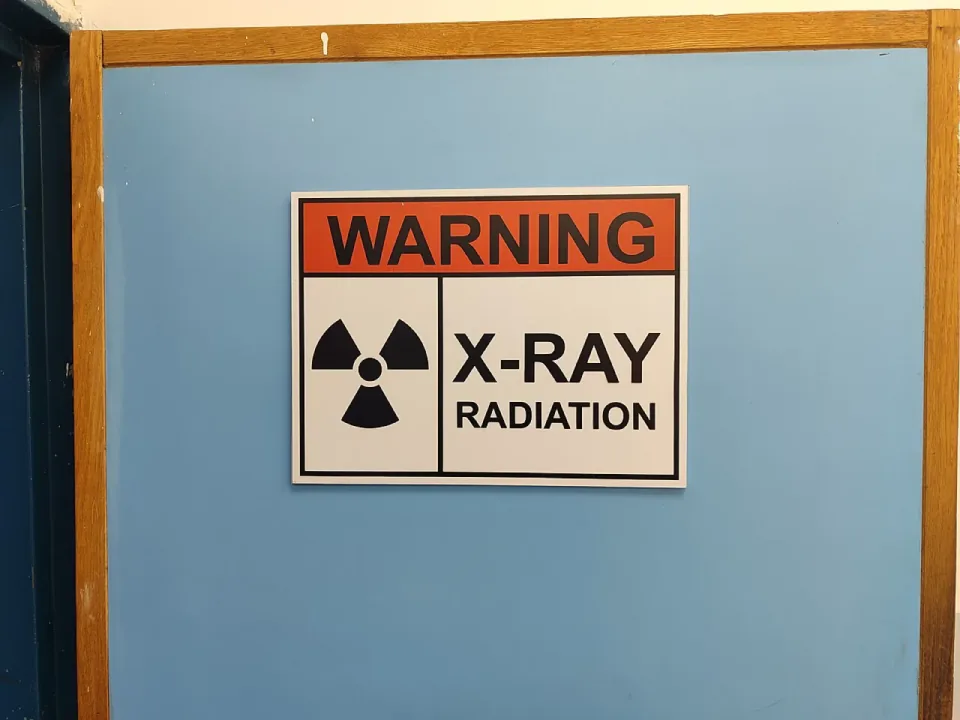
A patient cannot keep a receptor in place, and someone will have to hold it during the exposure. On who that should be, FDA directs that:

WARNING X-RAY RADIATION sign with the black radiation trefoil symbol mounted on the blue door of a radiography room
Image: أمين · CC BY-SA 4.0 · source

Tap an answer

Answer: A. Dental staff should not be the ones holding the receptor holder

Holding a receptor during an exposure puts a hand, and often a body, into or beside the primary beam. Doing it repeatedly is what turns an occasional problem into a routine occupational dose, so the direction is that the people who work in the room are not the ones who do it. A holding device is what should be reached for instead, and only in extraordinary circumstances does the task fall to someone accompanying the patient.

Source: The Selection of Patients for Dental Radiographic Examinations, U.S. Food and Drug Administration; section “RECEPTOR HOLDERS”. U.S. government work, public domain.

Question 9

Infection Prevention and Control

Loading the next plate, the assistant notices a smear of dried saliva on the plate-entry slot of the PSP scanner. What is the correct response?

Tap an answer

Answer: C. Stop, and clean and disinfect the contaminated part of the scanner by the method its manufacturer specifies before any more plates go through

The entire point of transporting and handling exposed receptors aseptically is to keep the reading equipment clean, so contamination found on the scanner has to be dealt with before it is handed on to every plate that follows. The manufacturer specifies the method because a scanner is high-technology equipment that many disinfectants, and any excess liquid, will damage - which is why CDC directs that manufacturers be consulted about protection of the associated computer hardware. Dry-wiping only moves the soil around, and deferring the clean-up to the end of the day contaminates the rest of the day's plates.

Source: Guidelines for Infection Control in Dental Health-Care Settings, 2003, Centers for Disease Control and Prevention (MMWR 52, RR-17); section “Radiology”. U.S. government work, public domain.

Question 10

Infection Prevention and Control

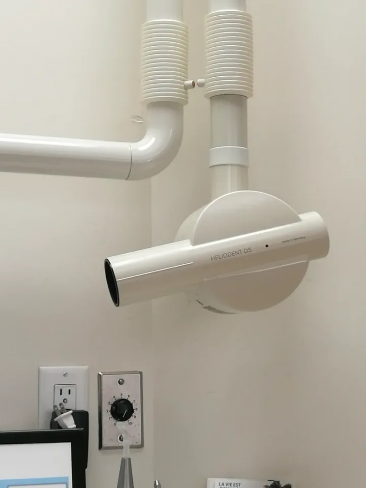
Pictured is a wall-mounted intraoral x-ray unit with its extension arm and round position-indicating device. Which pictured part most needs a disposable surface barrier before the patient is seated?

Wall-mounted intraoral dental x-ray unit with extension arm and round position indicating device
Image: Ptrump16 · CC BY-SA 4.0 · source

Tap an answer

Answer: D. The tubehead and position-indicating device that gloved hands grip while aiming the beam

Barriers go where gloved hands go. The tubehead and the position-indicating device are gripped and repositioned for every exposure by gloves that have been in the patient's mouth, which makes them clinical contact surfaces needing a fresh barrier for each patient. Parts that hands never reach are treated differently: the tube is sealed inside the housing, the wall bracket sits above head height, and the back of the arm is not handled during the series, so routine housekeeping is enough for all three.

Source: Guidelines for Infection Control in Dental Health-Care Settings, 2003, Centers for Disease Control and Prevention (MMWR 52, RR-17); section “Radiology”. U.S. government work, public domain.

Answer every question to see your score and how it splits by domain.

400 questions in the app

Keep going in Dentalingo

The other 390 questions for the DANB® RHS® exam, with the same explanations, lessons across its three domains, and a mock exam of 75 questions in 60 minutes.

The first 7 questions are free. After that, full access is a Study Camp subscription; the App Store shows the price in your country. More about the app

Download on the App Store
  • Dentalingo's Practice screen: daily quests and the questions due for spaced-repetition review.Spaced review of what you missed
  • Dentalingo's question screen: a chairside question about a blood pressure reading, answered correctly, with the explanation below the options.Answers explained the moment you choose
  • Dentalingo's Stats screen: answers graded, XP per day and an accuracy trend.XP and accuracy over time

Think an answer is wrong? Write to us with the question number and the page; every question is checked against its source, and a confirmed error is corrected in the app as well.