 Dentalingo
Question 1
Evaluation
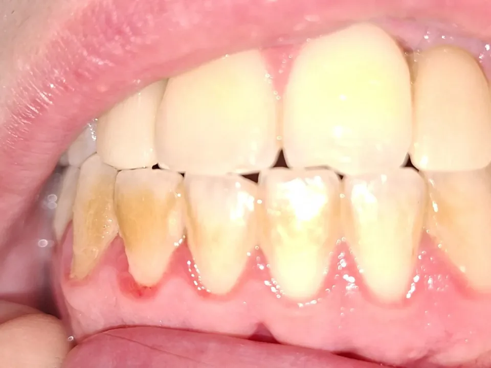
The photograph shows the front teeth of a patient being examined for gum disease, with heavy deposit along the gumline. Which of these findings can a radiograph add that this photograph cannot show?
Tap an answer
Answer: D. Loss of the bone supporting the teeth in that region
A photograph shows what is on and around the crowns: deposit, staining and the colour and contour of the gum margin. What it cannot show is what has happened to the supporting bone underneath, and that is what a radiograph adds — an x-ray image of the teeth and mouth can show whether bone has been lost as a result of gum disease. Pocket depth is measured with a probe, not read off any image, and the redness and the hard deposit are both already visible in the photograph itself.
Source: Periodontal (Gum) Disease, National Institute of Dental and Craniofacial Research; section “Diagnosis”. U.S. government work, public domain.