
Skip to content

Free practice test · from 320 questions

Anatomy, Morphology and Physiology practice test, every answer explained

A free sample from Dentalingo’s 320 questions for the DANB® AMP exam. Tap an answer to see at once whether it’s right, why, and the source it was checked against. The other 310, a timed mock exam and spaced review are in the app.

The format and domains are in the Anatomy, Morphology and Physiology exam guide; how the questions are checked is on the sources page.

Question 1

Head, Neck and Oral Cavity Anatomy and Physiology

This skull has been cut down the midline and is seen from the inside. On the floor of the cranium a saddle-shaped hollow is labelled, and the bone it is sunk into is named just beside it. Which bone carries that hollow, and what lies in it?
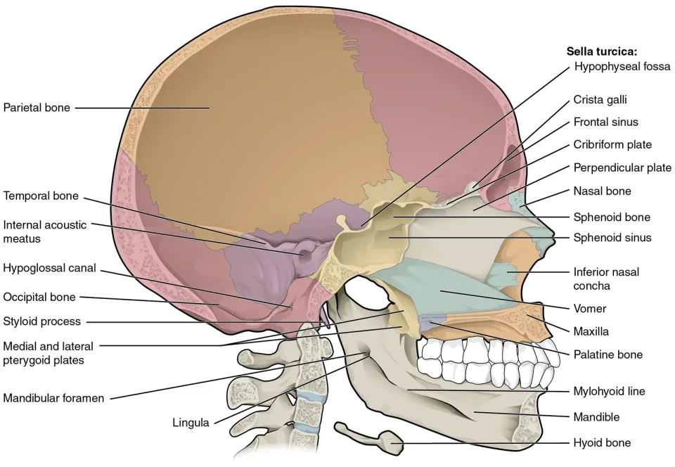
Colour-coded drawing of a skull cut down the midline and seen from inside. Towards the middle of the cranial floor a saddle-shaped hollow is labelled sella turcica, with its hypophyseal fossa, and it is sunk into the upper surface of the sphenoid bone, which is named just beside it. The frontal and sphenoid sinuses, the crista galli, the cribriform and perpendicular plates, the vomer, palatine bone, maxilla, inferior nasal concha, the pterygoid plates and the mandible with its foramen and lingula are labelled as well.
Image: OpenStax College, Anatomy & Physiology (Connexions), 2013 · CC BY 3.0 · source

Tap an answer

Answer: C. The sphenoid, on its upper aspect, cradling the pituitary gland

The saddle sits on the upper surface of the sphenoid bone and holds the pituitary gland. The sphenoid is the central bone of the cranial base and it also props the upper jaw from behind, which is why it turns up so often in head anatomy. The occipital carries the foramen magnum rather than a gland; the temporal bone holds the organs of hearing and balance deep inside its substance rather than in a socket on its surface; and the ethmoid's business with smell is a nerve passing through it.

Source: Dental Anatomy and Physiology (subcourse MD0501), U.S. Army Medical Department Center and School; section “2-3. CRANIUM”. U.S. government work, public domain.
Question 2

Head, Neck and Oral Cavity Anatomy and Physiology

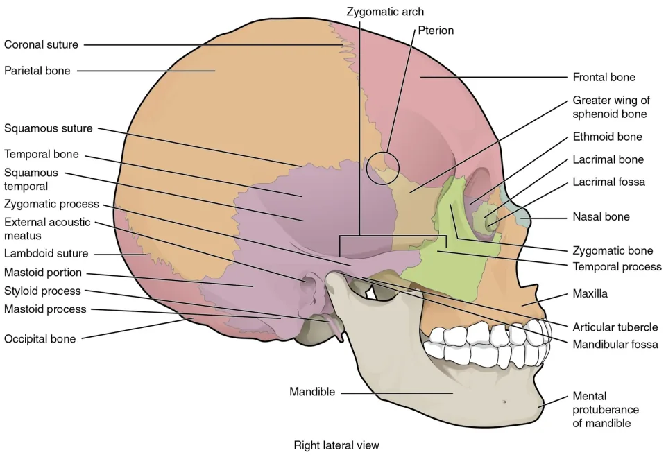
A bracket in this drawing of the skull from the side marks off the bar of bone running forward from in front of the ear to the cheek, and the two pieces that meet to make it are separately named on the drawing. Which two contributions make it up?

Colour-coded drawing of the skull from the right side, each bone in its own tint. A bracket labelled zygomatic arch spans the bar of bone running forward from in front of the ear to the cheek. Its back half is labelled as the zygomatic process of the temporal bone and its front half as the temporal process of the zygomatic bone, and the zygomatic bone itself is named beside it. The frontal, parietal, temporal, occipital and sphenoid bones, the maxilla and the mandible, the mastoid and styloid processes and the external acoustic meatus are labelled around them.
Image: OpenStax College, Anatomy & Physiology (Connexions), 2013 · CC BY 3.0 · source

Tap an answer

Answer: D. The zygomatic bone meeting the zygomatic process of the temporal bone

Two pieces meet end to end to complete the arch: the zygomatic bone of the face, which also makes the lower and outer rim of the orbit, and a projection running forward from the side of the temporal bone above the ear canal. The mastoid process of that same temporal bone points downward behind the ear instead, and the coronoid process belongs to the ramus of the lower jaw and lies beneath the arch rather than in it. The manual adds that this temporal projection is often caught up in fractures of the face.

Source: Dental Anatomy and Physiology (subcourse MD0501), U.S. Army Medical Department Center and School; section “2-4. FACE”. U.S. government work, public domain.
Question 3

Head, Neck and Oral Cavity Anatomy and Physiology

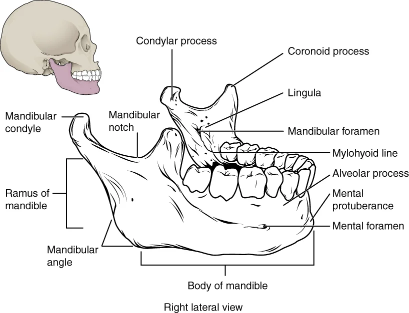
The upright part of this lower jaw ends above in two projections with a notch between them, and both are named on the drawing. Which of the two is the front one, nearer the teeth, and what attaches to it?

Line drawing of a lower jaw on its own seen from the right, the teeth towards the right of the frame, with a small skull inset showing where it sits. The upright ramus ends above in two projections separated by the mandibular notch: the coronoid process at the front, nearer the teeth, and the condylar process carrying the mandibular condyle behind it. The ramus, the body and the angle, the alveolar process, the mylohyoid line, the mandibular foramen and lingula, the mental protuberance and the mental foramen are labelled too.
Image: OpenStax College, Anatomy & Physiology (Connexions), 2013 · CC BY 3.0 · source

Tap an answer

Answer: B. The coronoid process, in front, taking the temporalis muscle

The front projection on the ramus is the coronoid process, and the temporalis takes its attachment there. The projection behind it is the condyloid process, which ends in the semi-cylindrical head called the condyle; that head, and not the coronoid process, works against the mandibular fossa. Between the two lies a U-shaped gap, the mandibular or sigmoid notch. The mastoid process is not on the lower jaw at all, but hangs from the temporal bone behind the ear.

Source: Dental Anatomy and Physiology (subcourse MD0501), U.S. Army Medical Department Center and School; section “2-7. THE LOWER JAW (MANDIBLE)”. U.S. government work, public domain.
Question 4

Head, Neck and Oral Cavity Anatomy and Physiology

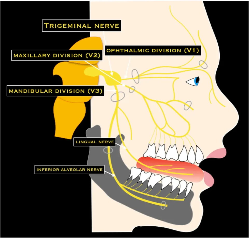
The trigeminal nerve is drawn in yellow on a head in profile here, and its three divisions are named where it leaves the brainstem. The middle one of the three fans out over the upper jaw and sends fine twigs down to the teeth in it. Which statement matches the account given of that division?

Schematic of a head in profile on a black ground with the trigeminal nerve drawn in yellow. Three divisions are named where the nerve leaves the brainstem: the ophthalmic division V1, running forward to the forehead and the eye; the maxillary division V2, which fans out over the upper jaw and sends fine twigs down to the upper teeth; and the mandibular division V3 below them. The lingual and inferior alveolar nerves are labelled lower down.
Image: Romano N, Federici M, Castaldi A — Insights into Imaging 10:33 (2019) · CC BY 4.0 · source

Tap an answer

Answer: A. It serves every upper tooth and carries no somatic motor fibres

The second division covers the middle of the face: the maxillary bone, every maxillary tooth, the soft tissue of the hard palate and the soft tissue around the teeth, and it is stated to have no somatic motor fibres at all. Motor fibres for chewing travel with the third division, and the soft tissue around the upper teeth is included, not excluded.

Source: Dental Anatomy and Physiology (subcourse MD0501), U.S. Army Medical Department Center and School; section “2-10. NERVE SUPPLY OF THE JAWS AND TEETH”. U.S. government work, public domain.
Question 5

Tooth Anatomy, Morphology and Physiology

When the arches meet normally, the upper front teeth both stand out past the lower ones and cover part of them. One term is used for the front-to-back projection and one for the up-and-down cover. Which term goes with which?

Tap an answer

Answer: B. Overjet is the front-to-back projection, and overbite the up-and-down cover

The manual splits the two overlaps by direction: the front-to-back one carries the name overjet and the up-and-down one the name overbite. Swapping them is the usual error. They are also not two readings of a single distance, and neither belongs to one arch by itself, because the overlap only exists as a relationship between the two arches.

Source: Dental Anatomy and Physiology (subcourse MD0501), U.S. Army Medical Department Center and School; section “4-2. ANATOMIC DIFFERENCES BETWEEN GROUPS OF TEETH”. U.S. government work, public domain.
Question 6

Tooth Anatomy, Morphology and Physiology

Four premolars are compared for size across the two arches. Which one is called the smallest of them?

Tap an answer

Answer: A. The lower first premolar, smallest of the four bicuspids

The smallest of the group is the first premolar of the lower arch. The first premolar of the upper arch is the opposite case, given the largest crown of its four. The lower second premolar is described as slightly larger and stockier than the lower first, not smaller.

Source: Dental Anatomy and Physiology (subcourse MD0501), U.S. Army Medical Department Center and School; section “4-21. MANDIBULAR FIRST BICUSPID”. U.S. government work, public domain.
Question 7

Tooth Anatomy, Morphology and Physiology

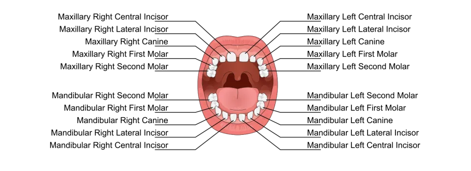
The same five kinds of tooth are named in each quadrant of this chart. How many of each kind does that come to?

Line drawing of an open mouth seen from the front, twenty primary teeth drawn in the two arches with wide spaces between them. Leader lines carry the full name of every tooth to the margins. Each quadrant lists five names and five only, in this order outward from the middle: central incisor, lateral incisor, canine, first molar, second molar. No premolar appears in any quadrant.
Image: Gregory david baker, Vectorization: Mrmw · CC BY-SA 4.0 · source

Tap an answer

Answer: C. Four of each kind, one per half-jaw right across the mouth

Two jaws with two halves apiece gives four of every kind, which is why naming a tooth needs both the jaw and the side before it identifies one tooth. Two would only cover a single jaw, five is the number of kinds rather than the number of each, and eight double-counts.

Source: Dental Anatomy and Physiology (subcourse MD0501), U.S. Army Medical Department Center and School; section “4-3. DECIDUOUS TEETH”. U.S. government work, public domain.

Question 8

Tooth Anatomy, Morphology and Physiology

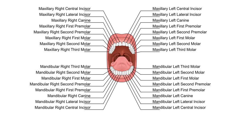
Ten teeth in each jaw of this chart stand in the places the first set once held. Which ten are they?

Line drawing of an open mouth seen from the front, all thirty-two permanent teeth drawn in the two arches. Leader lines run out to both margins and every tooth carries its full name in three parts: the arch, the side, and the tooth. Eight names run down each margin for the upper arch and eight for the lower. The names use the words canine and premolar rather than cuspid and bicuspid. The tongue and palate are drawn but not labelled.
Image: Gregory david baker, Vectorization: Mrmw · CC BY-SA 4.0 · source

Tap an answer

Answer: A. The ten standing furthest forward in each of the two jaws

Ten deciduous teeth per jaw are replaced by the ten permanent teeth nearest the front. All sixteen cannot be right, because the permanent molars have no predecessor at all; the ten furthest back reverses the group; and six would leave the two premolars on each side unaccounted for.

Source: Dental Anatomy and Physiology (subcourse MD0501), U.S. Army Medical Department Center and School; section “4-4. REPLACEMENT OF DECIDUOUS TEETH”. U.S. government work, public domain.

Question 9

Tooth Anatomy, Morphology and Physiology

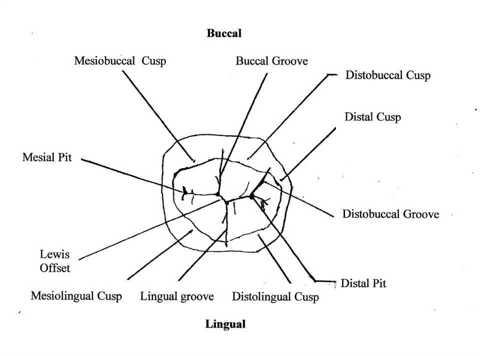
This drawing labels the biting surface of a lower first molar. In the normal relation a named cusp of the upper molar seats into one of these labelled features. Which one?

Hand-drawn outline of a lower first molar seen from its biting surface, Buccal printed at the top and Lingual at the bottom. Five cusps are labelled around the rim: Mesiobuccal, Distobuccal and Distal along the top and right, Mesiolingual and Distolingual along the bottom. Three grooves are labelled between them, Buccal Groove, Distobuccal Groove and Lingual groove, together with a Mesial Pit, a Distal Pit and one further label reading Lewis Offset.
Image: Acuteadvise · CC BY-SA 3.0 · source

Tap an answer

Answer: D. The buccal groove, labelled on the cheek side

The receiving feature is a groove and it lies on the cheek side, which is where the incoming cusp comes from. A pit is a point rather than a channel, a cusp is a peak rather than a channel, and the tongue-side groove faces the wrong way for an upper cusp to reach it.

Source: An Assessment of the Occlusion of the Teeth of Youths 12–17 Years, United States (Vital and Health Statistics, Series 11, No. 162), National Center for Health Statistics, 1977; section “1. Buccal segment relation”. U.S. government work, public domain.
Question 10

Tooth Anatomy, Morphology and Physiology

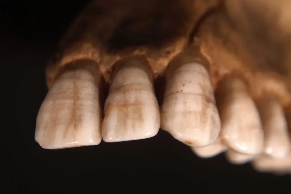
This dry specimen shows bands running across the front teeth. In hypoplasia, what has gone wrong with the enamel, and when?

Macro photograph of a dry upper jaw specimen against a black ground, the front teeth still standing in their bone. Across the outer surface of each front tooth run several narrow horizontal bands, parallel to one another and to the gum margin, stained a little browner than the enamel between them and set in rows down the crown. The biting edges are smooth and glossy and the bands do not reach them.
Image: Otis Historical Archives Nat'l Museum of Health & Medicine · CC BY 2.0 · source

Tap an answer

Answer: B. Its development is lacking or defective, and this happens before eruption

The fault is in how much enamel was laid down, and it happens while the crown is still forming inside the jaw. Anything that happens after eruption, whether acid or abrasion, removes enamel that was properly made rather than preventing it from being made.

Source: Oral and Maxillofacial Pathology (subcourse MD0511), U.S. Army Medical Department Center and School; section “2-14. HYPOPLASIA”. U.S. government work, public domain.

Answer every question to see your score and how it splits by domain.

320 questions in the app

Keep going in Dentalingo

The other 310 questions for the DANB® AMP exam, with the same explanations, lessons across its two domains, and a mock exam of 80 questions in 60 minutes.

The first 7 questions are free. After that, full access is a Study Camp subscription; the App Store shows the price in your country. More about the app

Download on the App Store
  • Dentalingo's Practice screen: daily quests and the questions due for spaced-repetition review.Spaced review of what you missed
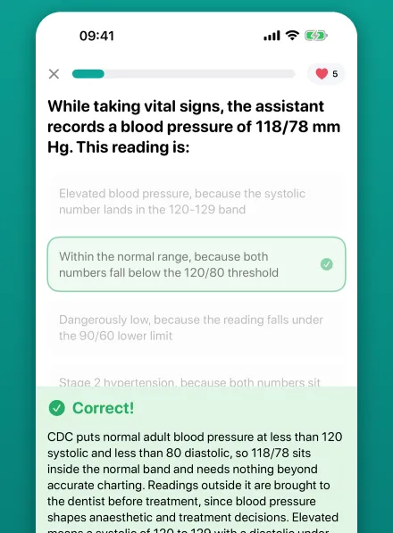
  • Dentalingo's question screen: a chairside question about a blood pressure reading, answered correctly, with the explanation below the options.Answers explained the moment you choose
  • Dentalingo's Stats screen: answers graded, XP per day and an accuracy trend.XP and accuracy over time

Think an answer is wrong? Write to us with the question number and the page; every question is checked against its source, and a confirmed error is corrected in the app as well.