
Skip to content

Free practice test 2 · from 320 questions

Anatomy, Morphology and Physiology practice test 2, every answer explained

Another free sample from Dentalingo’s 320 questions for the DANB® AMP exam, none of them from practice test 1. Tap an answer to see at once whether it’s right, why, and the source it was checked against. The other 310, a timed mock exam and spaced review are in the app.

The format and domains are in the Anatomy, Morphology and Physiology exam guide; how the questions are checked is on the sources page.

Question 1

Head, Neck and Oral Cavity Anatomy and Physiology

The gum in this drawing is shown lapping over the lowest part of the enamel, so that some of the enamelled surface would never be seen in the mouth at all. A chart entry records the clinical crown of a tooth. What exactly has been recorded?
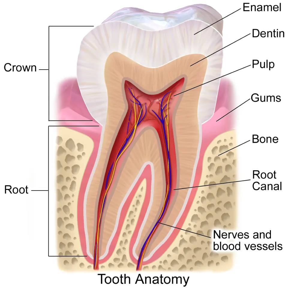
Colour illustration of a molar cut lengthways in the jaw. Enamel, dentin, pulp, the root canal and the nerves and blood vessels running in it are labelled on the tooth, with the gums and the surrounding bone named around it. The gum is drawn lapping over the lowest part of the enamel cap, so that part of the enamelled surface lies hidden beneath it, and the bracket marked crown reaches down behind the gum to the level where the enamel ends.
Image: BruceBlaus (Blausen Medical) · CC BY 3.0 · source

Tap an answer

Answer: B. The portion on show in the mouth, which rarely equals the enamelled part

Clinical crown measures what is exposed: whatever stands visible in the mouth on the day of the examination, and it seldom lines up with the area enamel covers. That enamelled area is the anatomic crown, a fixed quantity that does not change as gum tissue rides up or down the tooth.

Source: Dental Anatomy and Physiology (subcourse MD0501), U.S. Army Medical Department Center and School; section “3-5. PARTS OF TEETH”. U.S. government work, public domain.

Question 2

Head, Neck and Oral Cavity Anatomy and Physiology

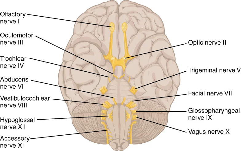
All twelve pairs of cranial nerves are named on this view of the underside of the brain. The fifth pair, drawn thicker than any of the others, leaves the side of the brainstem. That nerve has a motor root and a sensory root: what does each of the two carry?

Drawing of the underside of the brain with the cranial nerves picked out in yellow. All twelve pairs are named with their Roman numerals, from the olfactory nerve I at the front to the hypoglossal nerve XII at the back. The trigeminal nerve V is drawn thicker than any of the others and leaves the side of the brainstem.
Image: OpenStax, Anatomy & Physiology (OpenStax CNX) · CC BY 4.0 · source

Tap an answer

Answer: D. The motor root drives the muscles of mastication; the sensory root brings feeling in from the head and the face

The fifth nerve is mixed, and the manual splits the work cleanly: its motor root sends impulses to the chewing muscles while its sensory root brings sensation back from the structures of the head and face. Nothing there gives the nerve the muscles of the tongue or of the lips, and swapping the two roles around — sensation to the chewing muscles, movement from the face — is the commonest way this one is misremembered.

Source: Dental Anatomy and Physiology (subcourse MD0501), U.S. Army Medical Department Center and School; section “2-10. NERVE SUPPLY OF THE JAWS AND TEETH”. U.S. government work, public domain.

Question 3

Head, Neck and Oral Cavity Anatomy and Physiology

Nutrients and gases cross out of the blood into the surrounding tissue at one point in the circuit. Which vessel is that?

Tap an answer

Answer: A. The capillaries, the billions of tiny vessels that the arteries branch into

The exchange happens in the capillaries, the billions of tiny vessels the arteries branch into, because their walls are thin enough for food and oxygen to cross out of the blood and for carbon dioxide and waste to cross back in. Arteries and veins are transport vessels rather than exchange vessels: arterial walls are elastic and muscular, and venous walls, though thin, are collapsible and carry blood back to the heart behind paired valves. Lymphatic vessels drain lymph, not oxygenated blood.

Source: Dental Anatomy and Physiology (subcourse MD0501), U.S. Army Medical Department Center and School; section “1-9. CIRCULATORY SYSTEM”. U.S. government work, public domain.
Question 4

Head, Neck and Oral Cavity Anatomy and Physiology

The back third of the palate can be raised and lowered, while the front two-thirds cannot move at all. What accounts for the difference?

Tap an answer

Answer: D. The back third rests on no bone at all and is built mainly of muscle under its lining

What makes the front of the palate immovable is the bony plate beneath its covering; the back of the palate has nothing of the kind. It is a curtain of muscle clothed in mucous membrane, which is exactly why it can be lifted, and why its rear edge hangs free instead of ending against bone. Neither thinned bone nor cartilage is present there.

Source: Dental Anatomy and Physiology (subcourse MD0501), U.S. Army Medical Department Center and School; section “3-4. THE ORAL CAVITY”. U.S. government work, public domain.

Question 5

Head, Neck and Oral Cavity Anatomy and Physiology

A knob of bone can be felt jutting downward just behind the ear. What is it, and what does it belong to?

Tap an answer

Answer: A. The mastoid process, a projection of the temporal bone

The mastoid process is the part of the temporal bone felt as a knob behind the ear. It contains air cells that open into the middle ear and gives attachment to several muscles that move the head. The zygomatic process of the same bone runs forward from above the ear canal to meet the cheekbone, so it is felt in front of the ear instead. The coronoid and condyloid processes are the two projections on the upper edge of the ramus of the lower jaw, and neither is part of the cranium.

Source: Dental Anatomy and Physiology (subcourse MD0501), U.S. Army Medical Department Center and School; section “2-3. CRANIUM”. U.S. government work, public domain.

Question 6

Tooth Anatomy, Morphology and Physiology

Where do the vast majority of supernumerary teeth occur, and which is the most common?

Tap an answer

Answer: B. In the maxilla, the commonest being a mesiodens between the central incisors

Both halves of the answer come from one sentence: the upper jaw holds most of them, and the commonest sits in the midline between the upper central incisors. Moving it to the lower jaw, or naming a different extra tooth, breaks one half or the other.

Source: Oral and Maxillofacial Pathology (subcourse MD0511), U.S. Army Medical Department Center and School; section “2-18. SUPERNUMERARY TEETH”. U.S. government work, public domain.

Question 7

Tooth Anatomy, Morphology and Physiology

The lower first molar's two roots split from one another unusually high. How high, compared with other teeth?

Tap an answer

Answer: C. Closer to the crown than on any other tooth in either arch

This tooth's roots part company nearer the crown than the roots of any other tooth do, so the split is visible high up. Splitting near the tips, or at the midpoint like other molars, would put the division much lower, and the roots certainly do divide.

Source: Dental Anatomy and Physiology (subcourse MD0501), U.S. Army Medical Department Center and School; section “4-23. MANDIBULAR FIRST MOLAR”. U.S. government work, public domain.

Question 8

Tooth Anatomy, Morphology and Physiology

With the upper and lower teeth resting together, an examiner reads vertical overbite. Which part of the front teeth is compared?

Tap an answer

Answer: C. Their leading, or incisal, edges, one set against the other

The comparison is made at the cutting edges of the two sets of front teeth. A gum margin, a point of contact with a neighbour and a root tip are all real landmarks, but none of them is what this measurement is read from.

Source: An Assessment of the Occlusion of the Teeth of Youths 12–17 Years, United States (Vital and Health Statistics, Series 11, No. 162), National Center for Health Statistics, 1977; section “Vertical Overbite or Openbite”. U.S. government work, public domain.

Question 9

Tooth Anatomy, Morphology and Physiology

When a lower second premolar carries two cusps on its tongue side, what separates them?

Tap an answer

Answer: A. A groove running off the chewing surface and down the tongue side

A groove divides the pair and carries over the edge of the chewing surface onto the tongue side of the crown. A ridge joining the tips would fuse them rather than separate them, a central pit is a different feature, and the subcourse does describe a divider, so treating the cusps as meeting directly is wrong.

Source: Dental Anatomy and Physiology (subcourse MD0501), U.S. Army Medical Department Center and School; section “4-22. MANDIBULAR SECOND BICUSPID”. U.S. government work, public domain.

Question 10

Tooth Anatomy, Morphology and Physiology

How much of the chewing surface of a lower first premolar does its cheek-side cusp take up?

Tap an answer

Answer: B. About four fifths of it, leaving little for the tongue-side cusp

The cusp on the cheek side dominates the surface, taking roughly four fifths and leaving the tongue-side cusp very small, which is what gives this tooth its lopsided look. An even split, a reversed split, or a single cusp would all describe a different tooth.

Source: Dental Anatomy and Physiology (subcourse MD0501), U.S. Army Medical Department Center and School; section “4-21. MANDIBULAR FIRST BICUSPID”. U.S. government work, public domain.

Answer every question to see your score and how it splits by domain.

320 questions in the app

Keep going in Dentalingo

The other 310 questions for the DANB® AMP exam, with the same explanations, lessons across its two domains, and a mock exam of 80 questions in 60 minutes.

The first 7 questions are free. After that, full access is a Study Camp subscription; the App Store shows the price in your country. More about the app

Download on the App Store
  • Dentalingo's Practice screen: daily quests and the questions due for spaced-repetition review.Spaced review of what you missed
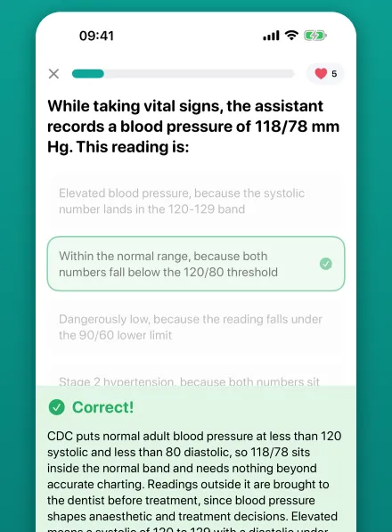
  • Dentalingo's question screen: a chairside question about a blood pressure reading, answered correctly, with the explanation below the options.Answers explained the moment you choose
  • Dentalingo's Stats screen: answers graded, XP per day and an accuracy trend.XP and accuracy over time

Think an answer is wrong? Write to us with the question number and the page; every question is checked against its source, and a confirmed error is corrected in the app as well.